Paper visualizing differently perfused anastomoses published in LIFE Journal
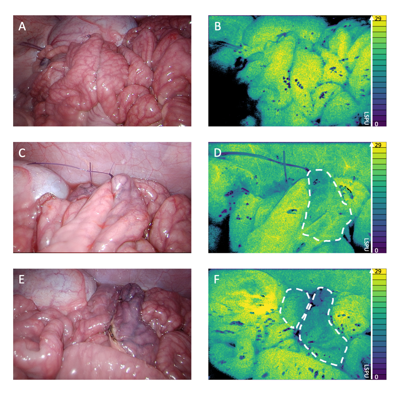
A new paper “Laparoscopic Laser Speckle Contrast Imaging Can Visualize Anastomotic Perfusion: A Demonstration in a Porcine Model ” published in August 2022 in cooperation with the group of prof. dr. Nicole Bouvy of MUMC highlights the significance of continuous real-time visualization of perfusion using PerfusiX-imaging, offering a new possibility for optimizing gastrointestinal surgeries. An Intestinal resection can lead to unavoidable vascular damage, which may not always be evident during intraoperative clinical assessments of local intestinal perfusion. Impaired perfusion, if left untreated, can lead to complications like anastomotic leakage (AL). To address this challenge, the researchers at the MUMC+ present a with PerfusiX-Imaging for perfusion assessment during anastomotic surgeries. The study involved isolating three segments from the small intestine of a pig while compromising the perfusion of each segment by coagulating several mesenteric arteries. Both clinical assessments and laser speckle imaging were employed to detect the induced perfusion deficits and subsequently guide a transection in either well-perfused, marginally perfused, or poorly perfused tissue areas within the segment. The researchers then created three differently perfused anastomoses: well-perfused/well-perfused, well-perfused/poorly perfused, and poorly perfused/poorly perfused. PerfusiX-Imaging played a crucial role by continuously assessing local intestinal perfusion and detecting perfusion deficits in real-time. The imaging feedback provided precise guidance during the surgical procedure. When evaluating the final anastomotic perfusion, LSCI demonstrated the ability to visualize varying degrees of perfusion accurately. In contrast, standard clinical assessment showed only minor differences in the visual appearance of the tissue.
The findings of the study underscore the significant potential of laparoscopic LSCI for intraoperative perfusion assessment. By providing real-time visualization of perfusion, this innovative technique offers a powerful tool for optimizing anastomotic surgeries. The ability to detect and address perfusion deficits promptly may eventually contribute to reducing the risk of complications, ultimately leading to improved patient outcomes. The paper is published open access and can be found by clicking here!