PerfusiX-Imaging
More clarity
for better clinical
decision making
How it works
Designed for specialists
Anastomotic leakage (AL) is a major complication worldwide with occurrence rates ranging up to 13% and a mortality of around 10%. AL occurs after a colon resection roughly 4 days post-surgery and requires re-operation with prolonged hospitalization. AL is multi-factorial, however the general consensus is that the state of perfusion at the site of the anastomosis is a major contributor. This is what Perfusix-Imaging aims to improve.
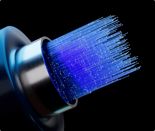
Laser Speckle Imaging
Currently surgeons determine the state of microcirculation based on experienced but subjective measures of viability, such as: tissue colour, palpable motion or bleeding at the resected edges. PerfusiX-Imaging is the development of a laparoscopic tool based on Laser Speckle Contrast Imaging (LSCI) that can visualize the state of microcirculation at any given time during laparoscopic surgery, without the need for injecting a fluorescent dye.
How it works
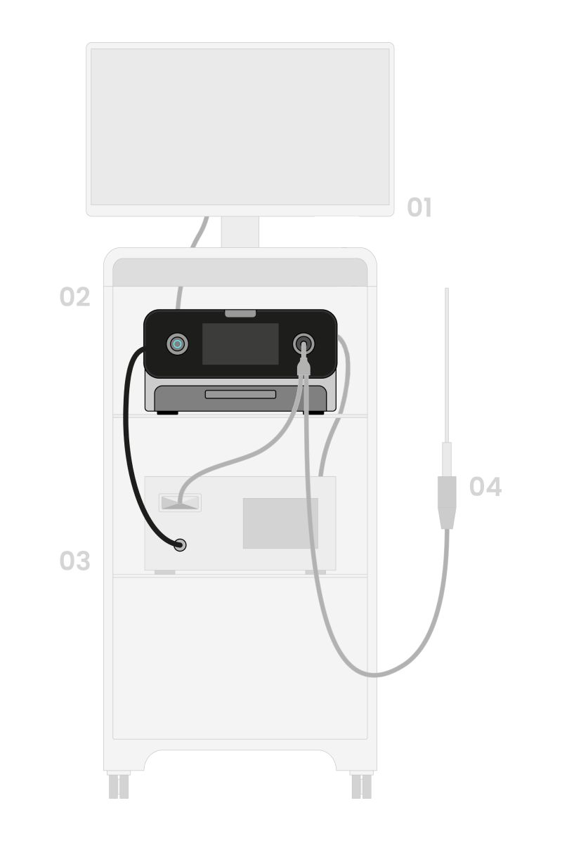
A traditional laparoscopy OR trolley consists of a laparoscopic device a monitor and the laparoscope. Perfusix-Imaging can be placed in the same trolley and be connected to the laparoscopy device. Perfusix-Imaging is connectable with most brands on the market.
01 Monitor
Displays the Real-time camera view or perfusion view.
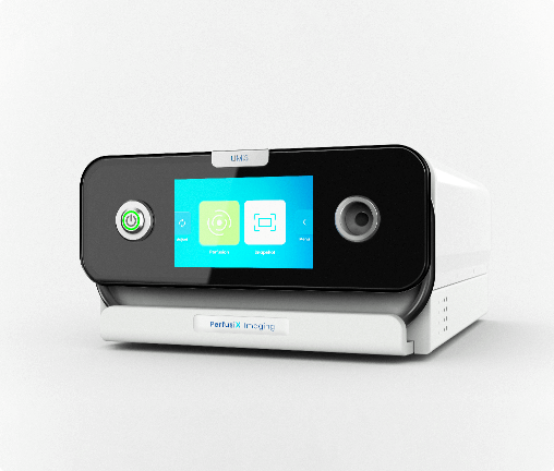
02 Perfusix-Imaging
Adds perfusion view to any laparoscopic device.
03 Video processor
Processes the images from the laparoscopic camera.
04 Laparoscope
The camera connected to the devices.
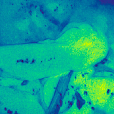
Identifying perfused tissue
Perfusix-Imaging is highly capable of distinguishing between perfused and non-perfused tissue. In multiple of our experiments the results are clear. They show us that we can not only visualize large local perfusion differences rapidly (under 1 second) but also reassure us of the added clinical value in identifying unwanted local perfusion deficits during surgical procedures.
Read our: latest newsFlexible and easy solution
Plug and play
Locate perfused tissue with the click of a button with our plug and play solution.
Cross compatible
Connect effortlessly to any Laparoscopy device using the universal connector.
Dye and waste free
Instantaneous visualization, without the use of a dye or disposables.
Display options
Adjust the view to your liking via the interactive interface with our presets.
Investing in (y)our future.
PerfusiX-IGS for new applications
Next to PerfusiX-Imaging we are working on another project, called the PerfusiX-IGS project. This project is focused towards developing an innovative surgical tool aimed at revolutionizing perfusion measurements in certain surgical fields. The PerfusiX-IGS project is an EFRO-subsidized project, funded by the EU, PerfusiX-IGS addresses the critical need for objective perfusion assessment in open surgeries prone to perfusion-related complications in multiple clinical needs. By integrating advanced imaging techniques, PerfusiX-IGS enables surgeons to accurately assess tissue perfusion levels, identify optimal surgical sites, and make informed decisions to minimize complications.
The project involves collaboration between LIMIS Development B.V. and the ZiuZ Group, and leading medical institutions including Medisch Centrum Leeuwarden (MCL), Universitair Medisch Centrum Groningen (UMCG), and Leids Universitair Medisch Centrum (LUMC). Through clinical validation and studies conducted with these partners, PerfusiX-IGS aims to validate dye free perfusion measurements in the clinics.
This project is co-financed by the European Union.